

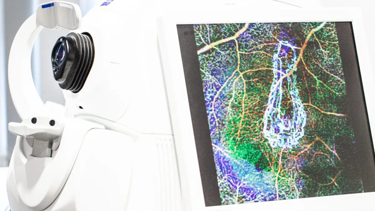
OCT of the optic disc
Optical coherence tomography (OCT) of the optic disc is a laser-based examination of the optic disc. This examination enables us to measure the optic nerve and the surrounding nerve fibres of the retina (retinal nerve fibre layer, or RNFL for short) with the utmost precision. If this examination is carried out regularly, it allows for what is known as a follow-up assessment.
What does the analysis show?
The results of each individual measurement are compared with values from a reference database. This enables us to carry out an individual risk assessment and determine whether glaucoma is present. In addition, the thickness of a specific layer of the macula – the ganglion cell layer – can be measured using optical coherence tomography. This is of great importance when it comes to detecting and monitoring the progression of early glaucomatous damage.
OCT angiography of the papilla
OCT angiography is a specialised examination. It provides a visualisation of the blood vessels in the optic disc and the vascular density of the surrounding retinal nerve fibre layer. Unlike fluorescein angiography, which is used to visualise the blood vessels of the fundus, this examination is carried out without the administration of a dye.
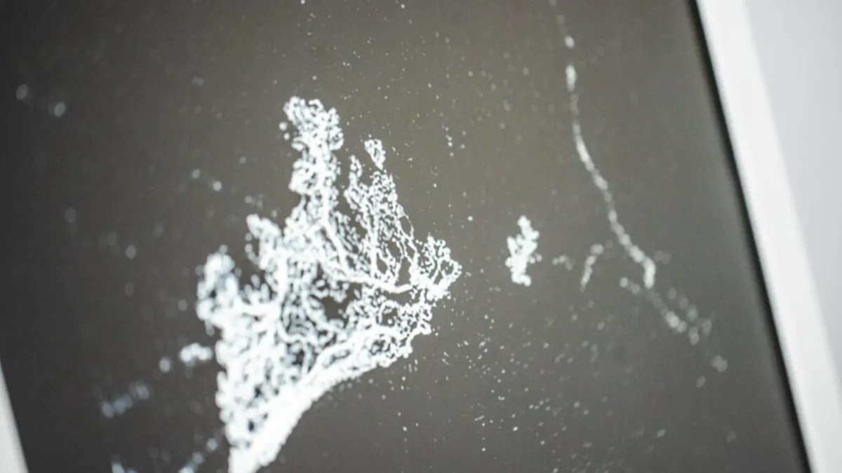
What does OCT angiography show?
OCT angiography measurements enable us to draw conclusions about the blood flow to the optic disc and the inner layers of your retina. In particular, comparing several images taken over time allows us to assess the stability of the findings.