OCT of the macula
In the case of macular diseases, we can use our treatment methods such as OCT (optical coherence tomography) to register precise indications of changes in the individual macular layers. This is important in order to be able to make an accurate diagnosis and thus carry out any necessary treatment and follow-up checks.
In diabetes, for example, retinal damage can lead to severe vision loss and even blindness. Optical coherence tomography is the most accurate method for measuring fluid deposits in the retina caused by diabetic changes. Moles at the back of the eye, the iris or the conjunctiva can also be precisely measured using OCT. This enables us to detect any changes or tumorous degeneration as early as possible. In addition to these examples, we can diagnose, measure and monitor many other changes in the macula using OCT.
OCT angiography
OCT angiography is a further development of the conventional examination for visualising vascular structures in the fundus of the eye. Several consecutive signals, which are scattered back from the red blood cells, the erythrocytes, are calculated over time. This allows the blood flow in the vessels to be visualised without administering a dye, as is the case with conventional fluorescein angiography. This visualisation of the vessels is used for wet macular degeneration, vascular occlusions or diabetic vascular changes. OCT angiography can also be used to visualise the vessels in the optic nerve.
The costs
If you have findings for which an OCT examination would be useful, we will inform you of this, as these examinations are not part of the statutory health insurance catalogue: The examinations are not included in the catalogue of services covered by statutory health insurance. They must therefore be billed according to the official scale of fees for doctors (GOÄ).
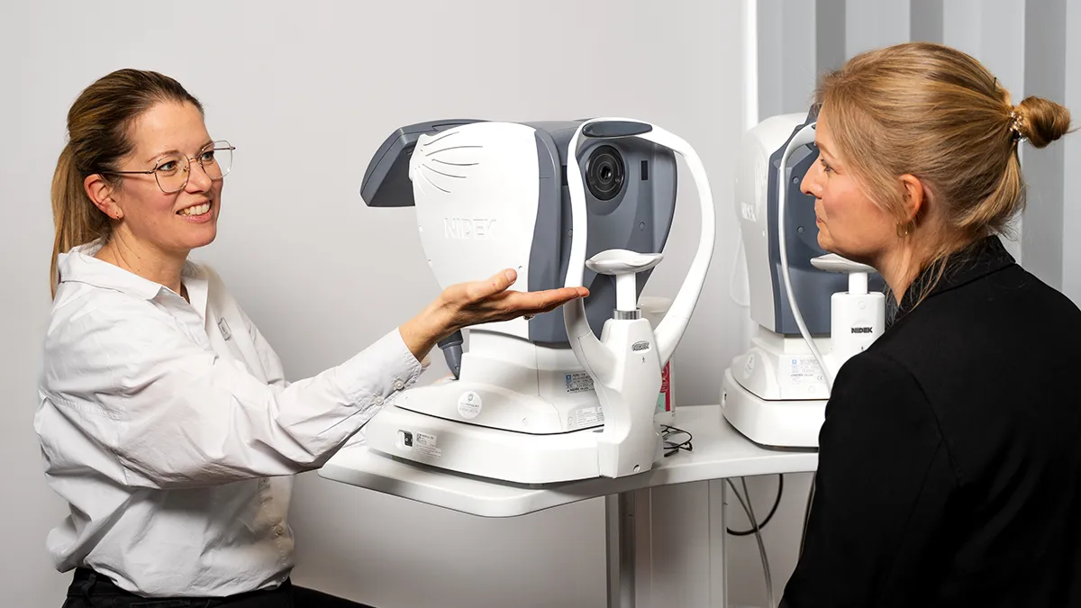
What is an OCT?
OCT is an abbreviation and stands for “optical coherence tomography”. This non-contact and painless examination enables changes to the optic nerve and retina to be diagnosed - in the shortest possible time and with maximum precision. This means that necessary treatments, operations and the effectiveness of administered medication can be precisely monitored. In detail, OCT technology enables the detection of pathological changes in the eye, such as glaucoma, diseases of the macula (the area of sharpest vision), diabetes or tumours. The accuracy of the examination is highly precise and therefore much more accurate than computer tomography (CT), magnetic resonance imaging (MRI) or ultrasound examinations.
Good to Know
What exactly is the macula?
The macula is the centre of the retina and is also known as the point of sharpest vision. The photoreceptors responsible for colour perception are present here in very high density. This enables a very high resolution of a visual impression.